
PNC-27: The Peptide That Targets Cancer Cells Without Harming Healthy Tissue
Exploring the Mechanism, Promise, and Research Behind the Selective Anti-Cancer Agent PNC-27
Researched and written by Keith Bishop, Clinical Nutritionist, Cancer Coach, Retired Pharmacist, and Founder of Prevail Over Cancer
PNC-27 and Cancer Introduction
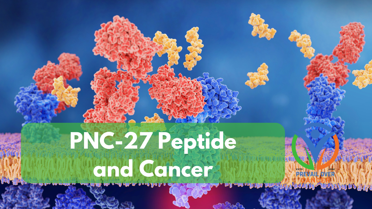
PNC-27 is an investigational anticancer peptide derived from the tumor suppressor protein p53. Unlike conventional chemotherapeutics that broadly target dividing cells, PNC-27 selectively binds to cancer cells expressing HDM-2 (also known as MDM2) on their outer membranes. This unique mechanism has sparked interest in its potential to induce rapid, necrotic cell death while sparing healthy tissue. Though not FDA-approved, PNC-27 represents a novel class of membrane-targeting peptides with implications for integrative oncology and terrain-based care.
PNC-27 Cancer Mechanism of Action
PNC-27 contains a p53-derived binding domain that targets HDM-2, a protein overexpressed on the surface of many cancer cells. Upon binding, PNC-27 inserts into the membrane and forms transmembrane pores (holes). This leads to necrosis (cell damage), rather than direct apoptosis (cell death), resulting in rapid cell rupture and the release of intracellular contents. Importantly, normal cells that lack HDM-2 membrane expression remain unaffected, highlighting the selectivity of this approach.[i] [ii] Furthermore, PNC-27 enters cancer cells and binds to the mitochondrial membrane, resulting in disruption.[iii]
PCN-27 Cancer Preclinical Evidence
PNC-27 has demonstrated potent cytotoxicity in vitro and in animal models:
- Breast Cancer ER+ PR+: PNC-27 induces breast cancer cell membrane lysis[iv]
- Colon cancer and colon cancer stem cells: PNC-27 kills colon cancer stem cells by binding of this peptide to membrane H/MDM-2. [v]
- Pancreatic cancer: Rapid necrosis and tumor regression[vi] [vii]
- Leukemia: Near-complete cell death, even in p53-deficient cells[viii] [ix] [x]
- Ovarian cancer: Selective killing of HDM-2–positive cells[xi] [xii] [xiii]
These studies underscore its potential across diverse tumor types, especially those resistant to apoptosis-based (cell death) therapies.
Cancers Expressing HDM-2
HDM-2 overexpression is a hallmark of many aggressive cancers. Here’s a list of tumor types with documented HDM-2 membrane expression:
- ✅ Pancreatic adenocarcinoma[xiv]
- ✅ Chronic myelogenous leukemia (CML) (AKA Chronic myeloid leukemia)[xv] [xvi]
- ✅ Mantle cell lymphoma[xvii] [xviii]
- ✅ Merkel cell carcinoma[xix]
- ✅ Multiple myeloma[xx] [xxi]
- ✅ Ovarian carcinoma (see above references)
- ✅ Head and neck squamous cell carcinoma (HNSCC)[xxii]
- ✅ Non-small cell lung cancer (NSCLC)[xxiii]
- Multiple cancers with documented HDM-2 overexpression[xxiv]
- ✅ Soft tissue sarcomas, especially liposarcomas
- ✅ Glioblastoma multiforme (GBM) - HDM2 contributes to p53 inactivation in a subset of tumors
- ✅ Breast cancer - Particularly in ER-positive subtypes
- ✅ Lung cancer - Non-small cell lung cancer (NSCLC) shows variable MDM2 expression
- ✅ Colon cancer - HDM2 overexpression linked to poor prognosis and p53 suppression
- ✅ Prostate cancer - HDM2 expression correlates with androgen-independent progression
- ✅ Osteosarcomas, soft tissue sarcomas, gliomas, lung carcinoma[xxv]
Cancers that May Not Express HDM-2
While HDM-2 (also known as MDM2) is frequently overexpressed in many aggressive cancers, particularly those with p53 pathway disruptions, its expression is not universal across all tumor types. Several cancers—including low-grade prostate cancer, clear cell renal carcinoma, papillary thyroid carcinoma, and certain indolent lymphomas—exhibit minimal or absent HDM-2 expression, especially on the cell membrane where targeted therapies like PNC-27 require access. This absence may reflect intact p53 signaling, low proliferative activity, or alternative oncogenic drivers. Importantly, most studies assess intracellular HDM-2 levels, not membrane localization, which is critical for therapeutic targeting. Diagnostic screening using flow cytometry or membrane-specific immunohistochemistry is essential to determine HDM-2 accessibility and avoid off-target or ineffective interventions. Understanding HDM-2 expression variability helps refine patient selection and underscores the need for personalized approaches in peptide-based cancer therapies.
Chart: Cancers with Low or Absent HDM-2 Expression
| Cancer Type / Subtype | HDM-2 Expression Status | Notes | Reference |
|---|---|---|---|
| Clear Cell Renal Cell Carcinoma | Low to absent | MDM2 expression is infrequent and correlates with higher tumor grade only | Haitel et al., Clin Cancer Res (2000) |
| Papillary Thyroid Carcinoma | Low | MDM2 knockdown reduces oncogenic activity and enhances iodine uptake | Shen et al., Cancer Genomics Proteomics (2025) |
| Low-Grade Prostate Cancer | Variable to low | MDM2 overexpression linked to higher grade and tumor volume; low-grade often lacks it | Leite et al., Mod Pathol (2001) |
| Hepatocellular Carcinoma | Low | MDM2–p53 axis dysfunction contributes to transformation; overexpression not universal | Meng et al., Cancer Res (2014) |
| Primary Melanoma | Low | MDM2 variants may influence risk and survival, but expression is generally low | Ward et al., Cancers (2023) |
| Indolent Non-Hodgkin Lymphoma | Low | MDM2 expression more common in aggressive subtypes; rare in indolent forms | Tzardi et al., Mol Pathol (1996) |
| IDH-Mutant Glioblastoma | Low | MDM2 amplification less common in IDH-mutant GBM; wild-type TP53 more frequent | Pellot Ortiz et al., Biomedicines (2023) |
| Testicular Germ Cell Tumors | Low | MDM2 overexpression linked to poor prognosis; wild-type p53 often retained | Lobo et al., Andrology (2020) |
- Membrane vs. intracellular HDM-2: Most studies assess total MDM2 expression, not membrane localization. PNC-27 requires membrane-bound HDM-2 for efficacy.
- Diagnostic screening: Flow cytometry or immunohistochemistry (IHC) with membrane-specific antibodies is essential before considering HDM-2–targeted therapies.
- Tumor heterogeneity: Even within a cancer type, expression can vary by grade, mutation status, or microenvironment.
PCN-27 Safety, Dosing, and Delivery
PNC-27 is not approved for human use and lacks standardized dosing. In vitro studies employed concentrations ranging from 10 to 500 µg/mL. Animal studies employed injections into tumors, but systemic (whole body) delivery remains experimental. No published Phase I human trials exist, and the safety of this treatment in humans is unknown.
Immune System Activation: What Happens When Cancer Cells Die
PNC-27 is a lab-designed peptide that targets a protein called HDM-2, which is often overproduced in cancer cells. When PNC-27 binds to this protein, it punches holes in the cancer cell membrane—causing the cell to rupture and die. This process is called lysis, and while it sounds like a victory, it’s only the beginning of a much larger immune response.
When cancer cells burst, they release a flood of internal materials—proteins, DNA fragments, and other cellular debris—into the surrounding tissue. These fragments act like distress signals, alerting the immune system that something unusual is happening. Specialized immune cells like macrophages and dendritic cells rush in to clean up the mess and present pieces of the dead cancer cells to other immune cells, essentially saying: “Here’s what the enemy looks like—go find more.”
This process can help the body recognize and attack remaining cancer cells. But if the cleanup is incomplete or overwhelmed, the immune system may become confused, exhausted, or even suppressed. That’s why supporting immune function during and after PNC-27 therapy is so important—it’s not just about killing cancer cells but helping the body process and respond to what comes next.
The dose of PNC-27 may have to be adjusted based on cancer cell dye off, immune response, and inflammation response. Work with your healthcare team to assess and adjust your program.
PNC-27 Anti-Inflammatory Synergy: Why Cleanup Matters
While immune activation is essential, it comes with a catch: inflammation. When cancer cells rupture, they release not only antigens but also inflammatory molecules that can irritate surrounding tissues. If this inflammation becomes excessive or chronic, it may:
- Damage healthy cells nearby
- Create a hostile environment that slows healing
- Distract the immune system from its cancer-fighting mission
- Trigger symptoms like fatigue, pain, or swelling
While PNC-27 shows promise in selectively targeting cancer cells, its effectiveness may be influenced by the body’s inflammatory and immune responses. Supporting these systems—through personalized nutrition, targeted supplementation, and integrative strategies—can help optimize cellular environments and enhance therapeutic outcomes. Because every patient’s biology is unique, it’s essential to work with a qualified healthcare provider to tailor these approaches.
PNC-27 Adjunctive Therapies and Terrain Support
To optimize outcomes and terrain resilience post-PNC-27 exposure, consider:
- Photobiomodulation: Enhances mitochondrial recovery and immune modulation
- Hyperbaric Normal Air Therapy: supports circulation and healing
- Hyperbaric oxygen therapy (HBOT): Supports tissue oxygenation and wound healing
- Sauna heat therapy: Promotes detoxification and heat shock protein immune activation
- Vagus nerve stimulation: Reduces inflammation and improves immune tone
- Supplements: to support circulation, inflammation, and the immune system
📘 Be the First to Know When the PNC-27 Book Launches
Curious about the science, protocols, and practical strategies behind PNC-27? My upcoming book dives deep into its mechanism, clinical relevance, and integrative support approaches. If you’d like early access and exclusive updates, you can join the wait list today. You’ll be notified as soon as the book is released—plus receive bonus content and early registration options for related webinars and coaching. Don’t miss your chance to stay ahead of the curve.
PNC-27 Cancer Controversies, Limitations, and Future Directions
Despite promising preclinical data, PNC-27 faces several hurdles:
- ❌ No human trials or FDA approval
- ⚠️ Risk of off-label use without safety data
- 🧪 Need for pharmacokinetic and immunogenicity studies
- 🔬 Unclear long-term effects on tumor microenvironment and immune memory
PNC-27 Sourcing
PNC‑27 is available only from research-grade peptide suppliers and custom peptide synthesis companies in the United States; it is sold strictly for laboratory research and not for human use. The FDA has warned consumers not to use PNC-27 products as cancer treatments because the products are unapproved and unverified, and are contaminated in tested samples.
Typical PNC-27 sources in the U.S.
- Research peptide vendors that sell lyophilized synthetic peptides for in vitro and animal research.
- Custom peptide synthesis companies that will manufacture PNC‑27 to order for investigators with an institutional research use agreement.
- Academic or commercial peptide core facilities that can synthesize or source PNC‑27 for an institutional laboratory.
PNC-27: What to expect and ask from suppliers
- Product format: lyophilized powder, variable purity grades (often specified by HPLC percentage).
- Labeling: “For research use only” or “Not for human or veterinary use.”
- You should ask for documentation: COA (certificate of analysis) detailing purity, sequence, and storage conditions.
- Shipping/handling: dry ice or cold‑chain for reconstituted material; MSDS and handling guidance for laboratory personnel.
PNC-27 safety, regulatory, and ethical considerations
- PNC‑27 is not FDA‑approved for the diagnosis or treatment of any disease; according to the FDA, using it in humans is illegal and unsafe.
- The FDA has issued explicit warnings about marketed PNC‑27 products promoted to cancer patients because of contamination and false therapeutic claims.
- Any acquisition or use should occur only within an authorized research setting under institutional review, biosafety oversight, and appropriate ethics approval.
PNC-27 Practical Guidance
- If you represent a research laboratory and need PNC‑27 for validated in vitro or approved animal studies, procure it from an established peptide supplier or a reputable custom synthesis provider and request a COA and batch-specific QC data.
- If you are a patient or caregiver seeking PNC‑27 as a therapy, speak with your oncology team about approved, evidence-based options and clinical trials. Some integrative practitioners may be willing to use this outside of traditional medicine.
Sources of PNC-27 for Research Purposes
Vendor list for PNC‑27 (research use only)
Request a copy of the Certificate of Analysis (COA) for the lot or batch purchased product.
PuraPeptides (PuraPeptides / Pura Labs)
PNC-27 Cancer Reference Sources
[i] Bowne, W. B., Adler, V., Sookraj, K. A., Wu, V., Shteyler, V., Patel, H., Oxbury, W., Zenilman, M. E., Michl, J., & Pincus, M. R. (2010). Anticancer peptide PNC-27 adopts an HDM-2-binding conformation and kills cancer cells by binding to HDM-2 in their membranes. Proceedings of the National Academy of Sciences, 107(5), 1918-1923. https://doi.org/10.1073/pnas.0909364107
[ii] Pincus, M. R., Silberstein, M., Zohar, N., & Bowne, W. B. (2024). Poptosis or Peptide-Induced Transmembrane Pore Formation: A Novel Way to Kill Cancer Cells without Affecting Normal Cells. Biomedicines, 12(6), 1144. https://doi.org/10.3390/biomedicines12061144
[iii] Sign In. Annclinlabsci.org. Published 2024. Accessed October 3, 2025. https://www.annclinlabsci.org/content/54/2/137.long
[iv] Sookraj, K.A., Bowne, W.B., Adler, V. et al. The anti-cancer peptide, PNC-27, induces tumor cell lysis as the intact peptide. Cancer Chemother Pharmacol 66, 325–331 (2010). https://doi.org/10.1007/s00280-009-1166-7
[v] Thadi A, Morano WF, Khalili M, et al. Molecular Targeting of H/MDM-2 Oncoprotein in Human Colon Cancer Cells and Stem-like Colonic Epithelial-derived Progenitor Cells. Anticancer Res. 2021;41(1):27-42. doi:10.21873/anticanres.14749 https://ar.iiarjournals.org/content/41/1/1
[vi] Krzesaj P, Adler V, Feinman RD, et al. Anti-Cancer Peptide PNC-27 Kills Cancer Cells by Unique Interactions with Plasma Membrane-Bound hdm-2 and with Mitochondrial Membranes Causing Mitochondrial Disruption. Ann Clin Lab Sci. 2024;54(2):137-148. https://pubmed.ncbi.nlm.nih.gov/38802154/
[vii] Bowne, W.B., Sookraj, K.A., Vishnevetsky, M. et al. The Penetratin Sequence in the Anticancer PNC-28 Peptide Causes Tumor Cell Necrosis Rather Than Apoptosis of Human Pancreatic Cancer Cells. Ann Surg Oncol 15, 3588–3600 (2008). https://doi.org/10.1245/s10434-008-0147-0
[viii] Thadi A, Lewis L, Goldstein E, et al. Targeting Membrane HDM-2 by PNC-27 Induces Necrosis in Leukemia Cells But Not in Normal Hematopoietic Cells. Anticancer Res. 2020;40(9):4857-4867. doi:10.21873/anticanres.14488 https://pubmed.ncbi.nlm.nih.gov/32878773/
[ix] Wang, H., Zhao, D., Nguyen, L. X., Wu, H., Li, L., Dong, D., Troadec, E., Zhu, Y., Hoang, D. H., Stein, A. S., Malki, M. A., Aldoss, I., Lin, A., Ghoda, L. Y., McDonald, T., Pichiorri, F., Carlesso, N., Kuo, H., Zhang, B., . . . Marcucci, G. (2019). Targeting Cell Membrane HDM2: A Novel Therapeutic Approach for Acute Myeloid Leukemia. Leukemia, 34(1), 75. https://doi.org/10.1038/s41375-019-0522-9
[x] Davitt K, Babcock BD, Fenelus M, et al. The Anti-Cancer Peptide, PNC-27, Induces Tumor Cell Necrosis of a Poorly Differentiated Non-Solid Tissue Human Leukemia Cell Line that Depends on Expression of HDM-2 in the Plasma Membrane of these Cells. Annals of Clinical & Laboratory Science. 2014;44(3):241-248. Accessed October 3, 2025. https://www.annclinlabsci.org/content/44/3/241.long
[xi] Anusha Thadi, Gleeson EM, Khalili M, et al. Anti-Cancer Tumor Cell Necrosis of Epithelial Ovarian Cancer Cell Lines Depends on High Expression of HDM-2 Protein in Their Membranes. Annals of Clinical & Laboratory Science. 2020;50(5):611-624. Accessed October 3, 2025. https://www.annclinlabsci.org/content/50/5/611.long
[xii] Alagkiozidis I, Gorelick C, Shah T, et al. Synergy between Paclitaxel and Anti-Cancer Peptide PNC-27 in the Treatment of Ovarian Cancer. Ann Clin Lab Sci. 2017;47(3):271-281. https://pubmed.ncbi.nlm.nih.gov/28667027/
[xiii] Ehsan Sarafraz-Yazdi, Gorelick C, Wagreich AR, et al. Ex vivo Efficacy of Anti-Cancer Drug PNC-27 in the Treatment of Patient-Derived Epithelial Ovarian Cancer. Annals of Clinical & Laboratory Science. 2015;45(6):650-658. Accessed October 3, 2025. https://www.annclinlabsci.org/content/45/6/650.long
[xiv] Bowne, W. B., Michl, J., Bluth, M. H., Zenilman, M. E., & Pincus, M. R. (2007). Novel peptides from the RAS-p21 and p53 proteins for the treatment of cancer. Cancer Therapy, 5B, 331. https://pmc.ncbi.nlm.nih.gov/articles/PMC2078333/
[xv] Peterson, L. F., Mitrikeska, E., Giannola, D., Lui, Y., Sun, H., Bixby, D., Malek, S. N., Donato, N. J., Wang, S., & Talpaz, M. (2011). P53 stabilization induces apoptosis in chronic myeloid leukemia blast crisis cells. Leukemia, 25(5), 761-769. https://doi.org/10.1038/leu.2011.7
[xvi] Donato, N. J., Fang, D., Sun, H., Giannola, D., Peterson, L. F., & Talpaz, M. (2010). Targets and effectors of the cellular response to aurora kinase inhibitor MK-0457 (VX-680) in imatinib sensitive and resistant chronic myelogenous leukemia. Biochemical Pharmacology, 79(5), 688-697. https://doi.org/10.1016/j.bcp.2009.10.009
[xvii] Richard J. Jones, Veerabhadran Baladandayuthapani, Sattva Neelapu, Luis E. Fayad, Jorge E. Romaguera, Michael Wang, Rakesh Sharma, Dajun Yang, Robert Z. Orlowski; HDM-2 inhibition suppresses expression of ribonucleotide reductase subunit M2, and synergistically enhances gemcitabine-induced cytotoxicity in mantle cell lymphoma. Blood 2011; 118 (15): 4140–4149. doi: https://doi.org/10.1182/blood-2011-03-340323
[xviii] Richard J. Jones, Qing Chen, Peter M. Voorhees, Ken H. Young, Nathalie Bruey-Sedano, Dajun Yang, Robert Z. Orlowski; Inhibition of the p53 E3 Ligase HDM-2 Induces Apoptosis and DNA Damage–Independent p53 Phosphorylation in Mantle Cell Lymphoma. Clin Cancer Res 1 September 2008; 14 (17): 5416–5425. https://doi.org/10.1158/1078-0432.CCR-08-0150
[xix] Houben R, Dreher C, Angermeyer S, et al. Mechanisms of p53 restriction in Merkel cell carcinoma cells are independent of the Merkel cell polyoma virus T antigens. J Invest Dermatol. 2013;133(10):2453-2460. doi:10.1038/jid.2013.169 https://www.jidonline.org/article/S0022-202X(15)35993-5/fulltext
[xx] Richard J. Jones, Chad C. Bjorklund, Veerabhadran Baladandayuthapani, Deborah J. Kuhn, Robert Z. Orlowski; Drug Resistance to Inhibitors of the Human Double Minute-2 E3 Ligase Is Mediated by Point Mutations of p53, but Can Be Overcome with the p53 Targeting Agent RITA. Mol Cancer Ther 1 October 2012; 11 (10): 2243–2253. https://doi.org/10.1158/1535-7163.MCT-12-0135
[xxi] Hurt EM, Thomas SB, Peng B, Farrar WL. Reversal of p53 epigenetic silencing in multiple myeloma permits apoptosis by a p53 activator. Cancer Biology & Therapy. 2006;5(9):1154-1160. doi: https://doi.org/10.4161/cbt.5.9.3001
[xxii] Hoffmann, T. K., Sonkoly, E., Hauser, U., Van Lierop, A., Whiteside, T. L., Klussmann, J. P., Hafner, D., Schuler, P., Friebe-Hoffmann, U., Scheckenbach, K., Erjala, K., Grénman, R., Schipper, J., Bier, H., & Balz, V. (2008). Alterations in the p53 pathway and their association with radio- and chemosensitivity in head and neck squamous cell carcinoma. Oral Oncology, 44(12), 1100-1109. https://doi.org/10.1016/j.oraloncology.2008.02.006
[xxiii] VanderBorght, A., Valckx, A., Van Dun, J. et al. Effect of an hdm-2 antagonist peptide inhibitor on cell cycle progression in p53-deficient H1299 human lung carcinoma cells. Oncogene 25, 6672–6677 (2006). https://doi.org/10.1038/sj.onc.1209667
[xxiv] Bowne, W. B., Michl, J., Bluth, M. H., Zenilman, M. E., & Pincus, M. R. (2007). Novel peptides from the RAS-p21 and p53 proteins for the treatment of cancer. Cancer Therapy, 5B, 331. https://pmc.ncbi.nlm.nih.gov/articles/PMC2078333/
[xxv] VanderBorght, A., Valckx, A., Van Dun, J., De Schepper, S., Vialard, J., Janicot, M., & Arts, J. (2006). Effect of an hdm-2 antagonist peptide inhibitor on cell cycle progression in p53-deficient H1299 human lung carcinoma cells. Oncogene, 25(50), 6672-6677. https://doi.org/10.1038/sj.onc.1209667
Author

Keith Bishop
Clinical Nutritionist, Cancer Coach, Retired Pharmacist, Author
Keith Bishop founded Prevail Over Cancer and is a passionate advocate for cancer awareness and research. With a background in pharmacy and clinical nutrition I’m dedicated to providing insightful and empowering information to help clients, survivors, and caregivers navigate their journey. From personal experiences and a commitment to holistic health, Keith aims to inspire and support the cancer community through comprehensive and accessible content.
Join over 8,000 subscribers!
Get access to Keith's Prevail Over Cancer Newsletter with news, blogs, and updates!
Recent Posts
Blog Post Topics
All Categories- microbiome modulation in oncology
25 hydroxyvitamin d cancer prevention
active hexose correlated compound
akkermansia and chemotherapy synergy
akkermansia and gastric cancer
akkermansia and radiation therapy
akkermansia muciniphila and cancer